Tagged: mushroom

#048: Mushroom Morphology: Jelly Fungi 0

#048: Mushroom Morphology: Jelly Fungi

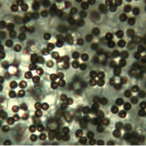
As you might guess, jelly fungi are distinguished by their gelatinous consistency. Their external appearance varies widely, so their texture is the only macroscopic feature that defines this group.  These fungi are placed within the phylum Basidiomycota, but they produce basidia (spore-bearing structures) unlike those of most other basidiomycetes (for more on basidia see FFF#013).  Because of this, they are often placed in the artificial group of fungi called heterobasidiomycetes.  The heterobasidiomycetes also include rusts and smuts, which do not form mushrooms.  Jelly fungi produce three different variations on the normal basidium (holobasidium) morphology.  Holobasidia have a bulbous, undivided base topped with spore-bearing steritmata.  The first variaition on this model is the cruciate basidium.  Cruciate basidia have a bulbous base divided into four cells by septa (cell walls).  The septa are at right angles to one another, making a cross shape when the basidium is viewed from above.  A good...

#047: Mushroom Morphology: Truffles 1

#047: Mushroom Morphology: Truffles

Truffles are ascomycetes that form below-ground (hypogeous) fruiting bodies. These mushrooms look like small, lumpy potatoes on the outside.  When cut open, truffles have a marbled appearance.  Like the false truffles and sequestrate basidiomycetes, true truffles have evolved to retain moisture in arid climates or other harsh conditions.  Truffles evolved from cup-shaped ascomycetes with a spore surface exposed to the air.  To keep the spores moist, the cup became closed.  Eventually, the interior surface became wrinkled and condensed, creating the marbled interior.  There are a number of truffle species that exhibit various stages along this evolutionary path.  Many of these truffles are hollow on the inside, as the gleba (interior, spore-producing tissue) has not fully become compacted.  All truffles rely on animals – usually small mammals – to dig up the fruiting bodies and eat them.  Truffles attract these animals by producing various scents.  These scents are what give edible...

#046: Mushroom Morphology: False Truffles 1

#046: Mushroom Morphology: False Truffles

False truffles are basidiomycetes that produce fruiting bodies at or below the soil surface (hypogeous). These mushrooms are included in the gasteromycetes because they develop their spores internally and have lost the ability to forcibly discharge their spores.  Like the true truffles, false truffles disperse their spores by attracting animals like insects, rodents, and deer.  False truffles secrete scents to attract these animals, which eat the mushrooms and spread the spores around in their feces.  Most false truffles have a gleba (the internal, spore-producing tissue) that is divided up into small chambers called locules.  Many false truffles also have a short stipe at the base or a stipe-columella that extends into the gleba as a branching structure.  Aside from the stipe-columella, the gleba of false truffles is fairly regular in appearance.  This easily separates them from true truffles, which appear marbled when sliced in half.

#045: Mushroom Morphology: Sequestrate Fungi 3

#045: Mushroom Morphology: Sequestrate Fungi

The sequestrate fungi* are an unnatural grouping of mushroom-forming basidiomycetes that have adapted to life in desert areas by keeping developing spores inside the fruiting body. Because of this they are included among the gasteroid fungi.  Like the other gasteromycetes, sequestrate fungi cannot form ballistospores (see FFF#013), and cannot forcibly discharge their spores.  Sequestrate fungi have a stipe and a cap-like head, similar to toadstools.  However, the head never fully opens and the spore-bearing surface remains enclosed by the cap.  The gleba (fertile tissue) does not develop regular gills or pores.  Instead, the gleba forms irregular pockets that are sometimes gill-like in appearance.  This is a highly diverse group of fungi, so there are many variations on this basic structure.  Some sequestrate fungi partially open their caps, while others never do.  Some have reduced stipes and are barely held above the ground.

#044: Mushroom Morphology: Earthstars 1

#044: Mushroom Morphology: Earthstars

In celebration of Independence Day, I will be discussing the festively-shaped earthstars. Earthstars are characterized by a puffball-like sphere surrounded by a star-shaped base.  The earthstars all have a three-layered peridium (the surface that covers the developing spore tissue).  At maturity, the two outer layers split into rays and peel back to form the star-shaped base.  The endoperidium (interior layer) then develops an ostiole (pore) ringed by a peristome through which spores are discharged.  Earthstars are included in the gastromycetes, which all form their spores internally and cannot forcibly discharge their spores.  Instead, like the puffballs, spores of earthstars are forced out of the pore when raindrops land on the endoperidium.  All earthstars have capillatum, a network of cells designed to help spores move to the pore when a raindrop strikes the mushroom.  If you cut open an earthstar you will find that it looks nothing like the interior of...

#043: Mushroom Morphology: Bird’s Nest Fungi 2

#043: Mushroom Morphology: Bird’s Nest Fungi

These small (< 1.5cm) fungi are easily recognizable by their striking resemblance to a bird’s nest with eggs inside. The “eggs” are actually spore-containing sacks called peridioles.  As in the other gasteromycetes, the bird’s nest fungi produce spores internally and have lost the ability to forcibly discharge their spores.  To overcome this obstacle, the bird’s nest fungi developed their unique morphology to act as a splash cup.  The basic splash cup mechanism works as follows: a raindrop falls into the cup, which propels one of the peridioles (“eggs”) out of the cup.  The next challenge for the bird’s nest fungi is to stick to whatever they land on.  These fungi have evolved two different mechanisms in order to accomplish this.  Fungi in the genera Mycocalia, Nidula, and Nidularia have peridioles that are covered in sticky mucilage, which allows them to adhere to surfaces on contact.  Fungi in the genera Crucibulum...

#042: Mushroom Morphology: Earthballs 2

#042: Mushroom Morphology: Earthballs

This group of mushrooms goes by a variety of common names, including “earthballs,” “earth balls,” and “false puffballs.” Additionally, all of these mushrooms belong to the family Sclerodermataceae and could casually be referred to as “sclerodermas.”*  Using these terms to distinguish earthballs from puffballs is a fairly recent development, so many earthballs are still commonly called puffballs.  Visually, immature earth balls can look similar to puffballs.  If you are collecting puffballs to eat, make sure you know how to tell the difference between true puffballs and the usually poisonous earthballs.  The main difference between earthballs and puffballs is that, unlike the puffballs, earthballs do not release their spores through a regular pore.  Instead, the peridium (outer layer) cracks and tears irregularly.  Like all gasteromycetes (fungi whose spores mature internally), earthballs have lost the ability to forcibly discharge their spores.  The spores mature in the center of the earthball, enclosed by...

#041: Mushroom Morphology: Puffballs 5

#041: Mushroom Morphology: Puffballs

The puffball is probably the second most familiar mushroom morphology. Many people can remember finding one of these as a child and giving in to the uncontrollable desire to kick the ball-shaped mushroom.  Anyone who has tried this knows that your efforts are rewarded with a fabulous puff of spores.  This puff is not just for the amusement of small children but is actually a rather ingenious spore dispersal mechanism.  Puffballs are gasteroid fungi, meaning that spores develop inside the mushroom.  All gasteroid fungi have lost the ability to forcibly discharge their spores, so they have to come up with other ways to release their spores.  Puffballs achieve this by puffing their spores out of small openings.  When the spores are mature a small opening (or a few small openings) called an ostiole develops on the surface of the mushroom.  When a raindrop (or an animal’s/ someone’s foot) lands on...

#040: Mushroom Morphology: Stinkhorns 2

#040: Mushroom Morphology: Stinkhorns

As the name suggests, this group is made up of the most pungent fungi. The defining characteristic of this group is that they all produce a dark, smelly slime that carries their spores.  This slime smells like rotting flesh for the express purpose of attracting flies and beetles.  The insects walk around in the spore slime and then fly away, carrying some of the mushroom’s spores on their legs.  After visiting a stinkhorn, the insects either land on real rotting material or on another stinkhorn.  In the first case, the rotting material serves as a substrate on which the spores can grow.  When the insect lands on another stinkhorn, this allows for cross-fertilization (much like insect pollination of flowers).  The other factor that all stinkhorns have in common is that they have a button stage.  All stinkhorns form a small, white, oval-shaped structure, or “egg” that contains the basic form...

#039: Mushroom Morphology: Crust Fungi 1

#039: Mushroom Morphology: Crust Fungi

This is a broad group of fungi that includes all basidiomycete mushrooms with an exposed spore surface that is smooth or mostly smooth. This group excludes all mushrooms that may be placed into the other morphological groups that I have described previously as well as all lichenized fungi.  For example, many chanterelles and corals have a mostly smooth hymenium but are not included in the crust fungi.  There are also a number of ascomycetes that fit this description, although they are usually not included at all in field guides.  These ascos tend to have small dots or pimples where the spores are released, whereas crust fungi do not have such a distinction.  Crust-like ascos also tend to be harder than their basidiomycete counterparts, making microscopic examination somewhat difficult.