#172: The Genus Amanita
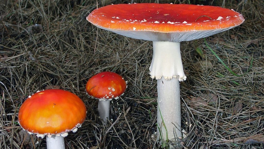
Amanita mushrooms, such as this A. muscaria, have a universal veil (note the warts on the pileus), partial veil (note the ring on the stipe), free gills, and a white spore print. By Chrumps [GFDL or CC BY-SA 3.0], via Wikimedia Commons (cropped)
Development
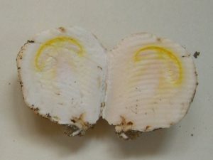
A. caesarea button showing developing pileus and gills. By Archenzo [GFDL], via Wikimedia Commons (cropped)
Universal Veil
The universal veil is produced as a result of this distinctive pattern of development. This veil comprises the outer layer of tissue that surrounds the button. In Amanita, the presence of a universal veil is an important marker for schizohymenial development. As the mushroom grows, it expands and eventually breaks through the universal veil.3
Universal veil remains usually take one of three forms: warts, patches, or volvas. In warts, the veil breaks up into small, regular fragments on top of the pileus. These fragments are pulled apart by the expanding pileus and become evenly spaced once the mushroom matures. In mushrooms that form patches, the universal veil does not break apart as easily. Instead, the veil remains in one irregularly shaped piece that sits on top of the pileus. If the mushroom forms a volva, the universal veil remains as a sac-like membrane surrounding the mushroom’s base.2,4
Unfortunately, universal veil fragments are frequently lost.1 Neither warts nor patches are attached to the pileus, so they slough off easily. Warts and patches often wash away in rain, brush off during collection or transportation, or simply disappear with age. Some amanitas have a powdery volva that is little more than a dusting on the dirt surrounding the mushroom base. Consequently, a mushroom could still be an Amanita even if you don’t find any remains of the universal veil.
Partial Veil
The partial veil covers the gills and extends from the stipe to the pileus margin. It serves to protect the developing gills and is cast off once the gills begin producing spores. Remains of the partial veil adorn either the stipe or the pileus margin. Partial veil fragments on the stipe of an Amanita usually take the form of a membranous ring. The ring may be large and skirt-like, as in A. muscaria, or may be little more than a thin band of tissue. On the pileus margin, partial veil pieces may hang down as irregular fragments of tissue.2,4 As with the universal veil, the partial veil remnants tend to be fragile and are often lost due to mechanical forces. Additionally, there are some amanitas that never leave partial veil fragments.1,2
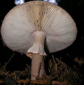
Amanita rubescens showing off its free gills and partial veil. By Danny Steaven S. [CC BY-SA 3.0], via Wikimedia Commons
Gills
Amanita species produce mushrooms with free gills. That is, the gills do not touch the stipe.2 In some mushrooms, this feature is very obvious because of a distinct gap between where the stipe ends and where the gills begin. However, in my experience it is rarely so clear-cut. I find that Amanita gills often touch the stipe, but do so only slightly. Thankfully, this is never enough to call them “attached.”
One feature that all Amanita species share is that they produce a white spore print.1,2 To take a spore print of an Amanita, cut off the stipe and put the cap gill-side down on non-white paper. Place a bowl or box over the cap to reduce air currents and retain moisture. Wait for an hour or two or until enough spores have accumulated to tell what color they are.
Microscopic Features
The features described above are helpful for identification of amanitas in the field, but their inconsistencies mean that they are not as useful for writing a description of the genus Amanita as a whole. Instead, mycologists define the genus using microscopic characteristics.
The family Amanitaceae (which includes the genus Amanita) is characterized by mushrooms that produce certain tissues in their gills and stipes. All Amanitaceae have a common arrangement of hyphae in their gills. When you slice a gill from top to bottom perpendicular to the long axis (so that the slice makes a V shape), you can see that the hyphae run straight down in the middle of the gill but run outward and downward toward each face of the gill. Amanitaceae mushrooms also produce large internal stipe tissue with large, club-shaped cells that are oriented vertically.5
These features are very robust and can still be identified after being cooked and partially digested. This is important for mushroom poisoning victims, since it allows an expert to tell whether the patient is likely suffering from amatoxin (FFF#091) poisoning.5
Amanita mushrooms are further differentiated by their schizohymenial growth. This feature is best observed in the button stage, but can be inferred from microscopic examination. In Amanita, the edges of the gills are adorned with irregularly shaped, bulbous cells and completely lack the smaller, spore-producing basidia (basidia are still present on the gill faces). The bulbous cells are formed as the gill edges separate from one another and the surrounding tissues. Other agarics have basidia on the gill edges because the gills do not tear apart from other tissues. Consequently, the lack of basidia on the edges of gills can be used as a marker for schizohymenial growth.3
Ecology
Another common feature of Amanita species is that they are mycorrhizal.2 Mycorrhizal fungi associate with plant roots, where they exchange nutrients from the soil for sugars the plant produced through photosynthesis. As a result, amanitas are almost always found under trees. Most Amanita species stick to certain types of trees, but they also tend to be flexible in their choice of hosts.2
Sequestrate Amanitas
A few species of Amanita do not fit the above descriptions at all. Instead of forming umbrella-like mushrooms, they form structures reminiscent of truffles or stalked puffballs. These species evolved alternate methods of spore dispersal and keep spores internally (“sequestrate”) instead of dropping them into the air currents. As a result, they no longer look like classic amanitas. However, the above-ground sequestrate forms still have a reduced form of a stipe. This stipe or “columella” still contains the vertical, club-shaped cells typical of Amanita species. This demonstrates that they do belong to Amanita despite their unusual morphology. Underground sequestrate Amanita species do not retain a stipe, so DNA evidence was used to place them into the genus Amanita.5
Tips for Identifying an Amanita
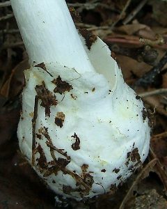
Volva at the base of Amanita virosa. By Jason Hollinger (Flickr: Destroying Angel) [CC BY 2.0], via Wikimedia Commons
Edibility
NEVER EAT AN AMANITA!
Edibility varies among amanitas. The genus includes the deadly poisonous A. virosa as well as the edible A. caesarea.1 Many people eat amanitas, but just as many avoid the entire genus to ensure they do not make a deadly mistake. Personally, I don’t eat amanitas. For one, I don’t trust myself to identify A. rubescens accurately enough. Even when I am sure of my identification, the words of a fellow mushroom hunter dissuade me from eating it: “No mushroom is good enough to risk dying from it.”
Online resources (including this site) usually recommend that people never eat Amanita species. This is probably to protect themselves from lawsuits, but it is also good advice. Because of the great diversity within the genus, you should never eat an Amanita until you are familiar with it and all of its look-alikes in your geographical area. The World Wide Web is a fantastic resource, but its global nature means that the information you find may not be applicable to your region. Local mushroom knowledge is more useful than the internet when it comes to edible amanitas.
Taxonomy
Amanitas belong to the core group of agarics (Agaricales). They share the family Amanitaceae with the genus Limacella. Limacella mushrooms are closely related to amanitas and have a very similar morphology. Species in Limacella can usually be differentiated by their slimy caps.2,6 For more on Limacella, see this page from Amanita expert and professional mycologist Rod Tulloss’ website, Studies in the Amanitaceae.
Amanita is a highly diverse genus, probably containing over a thousand species. Consequently, mycologists divided the genus into two subgenera, each with multiple sections.3,5 For a description of each Amanita section, see this page from Studies in the Amanitaceae.
| Kingdom | Fungi |
| Division | Basidiomycota |
| Subdivision | Agaricomycotina |
| Class | Agaricomycetes |
| Subclass | Agaricomycetidae |
| Order | Agaricales |
| Family | Amanitaceae |
| Genus | Amanita Pers.7 |
This post describes a group of mushrooms and as such the information on this page (including the pictures) cannot be used to identify any mushroom in particular.
This post does not contain enough information to positively identify any mushroom. When collecting for the table, always use a local field guide to identify your mushrooms down to species. If you need a quality, free field guide to North American mushrooms, I recommend Michael Kuo’s MushroomExpert.com. Remember: when in doubt, throw it out!
See Further:
http://www.amanitaceae.org/?Home
http://www.mushroomexpert.com/amanita.html
Citations
- Amanita. The Journal of Wild Mushrooming (2014).
- Michael Kuo. The Genus Amanita. MushroomExpert.Com (2013). Available at: http://www.mushroomexpert.com/amanita.html. (Accessed: 6th January 2017)
- RE Tulloss. About Amanita. Studies in the Amanitaceae (2017). Available at: http://www.amanitaceae.org/?About%20Amanita. (Accessed: 6th January 2017)
- Michael Kuo. Glossary of Mycological Terms. MushroomExpert.Com (2006). Available at: http://www.mushroomexpert.com/glossary.html. (Accessed: 6th January 2017)
- RE Tulloss. About the Amanita family. Studies in the Amanitaceae (2017). Available at: http://www.amanitaceae.org/?About%20Amanitaceae. (Accessed: 6th January 2017)
- RE Tulloss. About Limacella. Studies in the Amanitaceae (2017). Available at: http://www.amanitaceae.org/?About+Limacella. (Accessed: 6th January 2017)
- Amanita. Mycobank Available at: http://www.mycobank.org/BioloMICS.aspx?TableKey=14682616000000067&Rec=56034&Fields=All. (Accessed: 6th January 2017)

![#005: Xylaria polymorpha, Dead Man’s Fingers [Archived]](https://www.fungusfactfriday.com/wp-content/themes/hueman/assets/front/img/thumb-medium-empty.png)


![#011: Characteristics of Kingdom Fungi [Archived]](https://www.fungusfactfriday.com/wp-content/themes/hueman/assets/front/img/thumb-small-empty.png)
5 Responses
[…] Morphologically, lepiotoid mushrooms most closely resemble mushrooms from the genus Amanita (FFF#172). Lepiotoid mushrooms have free gills, a white spore print, and a partial veil, but are saprobic […]
[…] species can also cause some confusion with Amanita (FFF#172) mushrooms, especially when young. Spore color is the easiest way to separate the two genera: […]
[…] partial veils that are membranous, that is, the tissue is thick and coherent; species of Amanita (FFF#172), for example, often have thick partial veils that in maturity remain hanging off the stipe like a […]
[…] Amanita6-9 […]
[…] to be pared down quite a bit (see FFF#177). The Blushers definitely belong to the genus Amanita (FFF#172), although their exact placement within the genus may change in the future. Fortunately, that […]