#224: Pythium insidiosum, Pythiosis
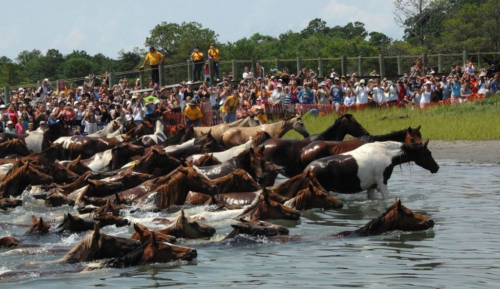
It’s a new year and already a fungus-like disease is making national headlines. Four wild ponies on Chincoteague were euthanized due to a disease called “Swamp Cancer” on December 28, 2018, bringing the total Chincoteague ponies killed by the disease in 2018 up to seven. Swamp Cancer is not a cancer at all; the disease is caused by the oomycete Pythium insidiosum. If you have a pet you may have heard of this disease before: P. insidiosum occasionally infects dogs and cats, often killing its hosts. Rarely, P. insidiosum causes disease in humans. P. insidiosum normally decays plant matter in flooded tropical to sub-tropical environments, so it is most often contracted in warm stagnant water. Because it reproduces only in those environments, P. insidiosum cannot spread from one animal to another.1–4
Pythium insidiosum
Pythium insidiosum is a fungus-like organism that belongs to the class Oomycota with the rest of the strange organisms known as “water molds.” These organisms are common in watery ecosystems and most infect plants or aquatic animals. P. insidiosum usually decomposes plant material but is unique among the water molds because it can also infect mammalian tissues.5
In its natural habitat, P. insidiosum lives in waterlogged areas like swamps, ponds, flooded yards, etc. and breaks down leaves and other plant debris in the water. P. insidiosum usually lives in tropical and sub-tropical areas, although its range has recently expanded into temperate regions.5,6
The life cycle of P. insidiosum starts as a spore swimming through the water. Like most oomycetes, the motile spores are powered by two flagella: one smooth “whiplash” flagellum and one fuzzy “tinsel” flagellum. These two types of flagella allow the spore to direct its movement and seek out nearby plant material.5
Once the spore finds a suitable substrate, it casts off its flagella and sticks to the substrate. The spore then germinates and sends out hyphae, which penetrate and begin decomposing the substrate. As the oomycete grows, it begins producing asexual spores. These spores are formed in a circular structure called a sporangium and develop two flagella. Once the spores mature, the sporangium ruptures and releases the spores into the water. Since the spores have flagella, they can swim through the water and find new areas to decompose.5
When resources run low, P. insidiosum may produce sexual spores called “oogonia.” Oogonia do not have flagella and cannot swim. However, oogonia have thick walls and are better equipped to survive adverse conditions. This is important for P. insidiosum, since it is often found in temporary bodies of water. When the water dries up, the oogonia still survive and can germinate to begin decomposition again once the area is flooded again.5
P. insidiosum is also able to infect animals. The spores will swim toward injured animal tissue, germinate, and send out infective hyphae. This is the same process used to infect plant material. However, the oomycete’s life cycle usually ends when it infects an animal. It cannot produce spores in animals, so the infection cannot be passed to other animals and the fungus cannot return to the swampy areas it normally inhabits. Once its host dies, P. insidiosum also dies.5
In horses, however, P. insidiosum forms hard structures called “kunkers.” Hyphae in kunkers have been found to produce spores if the kunkers are placed in water. Therefore, the fungus could complete its life cycle if one of the kunkers happened to fall out of an open wound and into standing water.5 I haven’t found any research into whether this happens naturally, so it is unclear if this is actually a way that spores return to their normal environment.
However, since P. insidiosum spores actively seek out animals to infect, it stands to reason that P. insidiosum can successfully return to the ground after infecting at least some animal(s). But why would P. insidiosum want to infect animals (especially when the chances of successfully completing its life cycle through an animal host seem to be extremely low)? Perhaps it’s a dispersal strategy: P. insidiosum could infect an animal in one area and then fall to the ground in another area. This would allow the oomycete to travel much longer distances than it could just by swimming through the water. Alternatively, this could be a survival strategy: P. insidiosum infections tend to last a long time, so the oomycete could survive a drought inside an animal and later return to the ground once conditions are more favorable. Obviously, this is all speculative and there are probably many other possible explanations. We really don’t know why this happens because most P. insidiosum research focuses on its role as a pathogen, not on how it survives in the wild. More research needs to be done to understand P. insidiosum’s ecological role and why the oomycete chooses to infect animals.
Disease
The disease caused by P. insidiosum – called “pythiosis” in any animal – is contracted when a spore enters the animal’s body. This most commonly happens when a spore swims to an open wound (such as a small cut) and attaches.5 P. insidiosum can also infiltrate the body when a spore is swallowed (common in dogs and cats around contaminated water).4,7 The course of the disease varies based on where the infection occurs as well as the infected organism.7
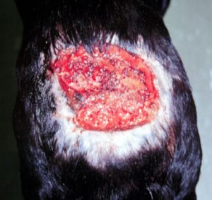
Horses contract an external form of the disease that is often called “swamp cancer.” After attaching to a wound, the spore germinates and the hyphae extend into the surrounding tissues. The hyphae kill the tissue and cause an open wound. The foul-smelling wounds begin small but quickly grow and exude a slightly bloody liquid. Sometimes they develop into large swollen areas that look cancer-like (thus the disease’s name). In severe cases, the wounds can cover large areas of a horse’s body, such as an entire leg. At the center of swamp cancer lesions, dead tissue condenses into hard yellowish branching structures called “kunkers.” Kunkers contain hyphae of P. insidiosum and are easy to remove from the wound. Blood and tissue loss from the open wound will leave horses anemic and eventually result in the horses’ death.6,7
Dogs can contract three forms of the disease: an external version, a gastrointestinal version, and a respiratory version. Like in horses, the external form of the disease causes open wounds that refuse to heal and fill with pus and blackened dead tissue. However, no kunkers form in dogs. The gastrointestinal form is the most common form of the disease in dogs and primarily affects younger adult dogs, especially Labrador Retrievers. This form usually causes inflammation in the small intestine or stomach, although infection can occur anywhere from the esophagus through the colon. Symptoms of gastrointestinal pythiosis include: vomiting, diarrhea, weight loss, anorexia, fever, and abdominal pain.7,8 The respiratory version is characterized by stuffiness, fever, and coughing.8 Any form can cause severe wounds that can lead to death if untreated.7
Pythiosis in cats is rare, but can occur. Cats most contract either an external form of the disease or a gastrointestinal version. Symptoms of pythiosis are generally the same as they are in dogs.7,9
Humans can also suffer from pythiosis, although this is rare. Pythiosis in humans is most common in places where humans frequently contact stagnant water, such as in areas suffering from floods. Infection primarily results in external wounds that do not heal. These open wounds can lead to other infections and in severe cases can result in death.10 In the United States, there have been several cases of children contracting the disease on their faces. It is unclear why this form of the disease is particular to the United States.11
Treatment
Three courses of treatment available are available for both external and internal forms of pythiosis: surgery to remove the infection, treatment with antifungal drugs, and “vaccination.”6–10
Surgery is the first line of treatment and can be used to treat any animal. Since P. insidiosum usually (at least before the late stages of the disease) causes a single localized infection, surgery can effectively remove the dead tissue as well as all the P. insidiosum hyphae.6–10 In dogs and cats, surgery is followed by using a laser to burn the surrounding tissue and kill any hyphae that were missed during the surgery.8,9 If the infection is discovered and identified early on and is still small, surgery is usually effective at clearing up the infection.7 Of course, the infection’s location is also important: P. insidiosum causing a skin ulcer will be easier to remove than an internal infection near a major artery.10
Antifungal drugs are used in conjunction with surgery and when the infection is in an area that is difficult to reach surgically.6–10 Since P. insidiosum is not a fungus, antifungal drugs are not particularly effective – only 20% of dogs respond to treatment with antifungal drugs.6,7 However, there are no specific drugs targeting oomycetes, so antifungals are currently the best option. Itraconazole is the first drug of choice, followed by terbinafine, and sometimes amphotericin B.7–10
In horses, dogs, and humans, another option is available: an immunotherapy “vaccine.”6–8 This treatment uses antibodies to encourage the animal’s own immune system to attack the P. insidiosum hyphae. Because it merely treats infection and does not prevent it, immunotherapy is not a true vaccine. The vaccine is administered by subcutaneous injection on one, seven, 21, and (if needed) 28 days after beginning treatment. In horses, the vaccine successfully cures 100% of cases less than 60 days old and 50% of long-term cases.7 Once the immunotherapy shrinks the infected area, surgery may be used to remove the remaining infection.7
The Chincoteague ponies were treated with surgery, immunotherapy, and antifungal drugs. This worked for one pony early in the year, but failed to beat the disease in later cases. Successful treatment was hampered by the fact that the horses are wild. Because they spend most of their time away from human eyes, infections tend to go unnoticed for a long time. This lowers the chances of successful treatment. With such a tragic ending to 2018, the Chincoteague Volunteer Fire Company (which manages the ponies on Chincoteague and the Virginia part of Assateague) will be more vigilant for Swamp Cancer in 2019 and hope spot the disease earlier on. They also hope to get access to a true vaccine that will prevent disease but is not yet approved by the FDA.1–3
Taxonomy
The taxonomy of the Oomycota (also written Oomycetes) has not been finalized. It was once placed in kingdom Fungi (and still is on the Integrated Taxonomic Information System), but has since been moved out.12,13 Its exact placement is uncertain. Some sources place the Oomycota as class in kingdom Stramenopila without specifying a phylum, but Wikipedia lists it as a class in phylum Heterokontophyta, which appears to belong to the kingdom Chromista.14–16 This reflects how little we actually know about this bizarre group of organisms. Even within the oomycetes, taxonomic relationships are not very well defined: Pythium is placed in the family Pythiaceae, but the order varies based on the source you’re consulting.14,16 The bottom line is that the oomycetes are poorly understood and need to be studied more. Yes, that will result in more taxonomic changes, but that’s better than the mess we have now.
Whatever the taxonomy, the oomycetes appear to be more closely related to certain algae than they are to fungi.15 How these organisms lost their ability to photosynthesize and developed a hyphal growth mode is sure to be a fascinating story that someone will hopefully figure out sometime.
| Kingdom | Stramenopila or Chromista (?) |
| Phylum | Heterokontophyta (?) |
| Class | Oomycota or Oomycetes |
| Order | Pytheales or Peronosporales |
| Family | Pytheaceae |
| Genus | Pythium |
| Species | Pythium insidiosum12,15,16 |
Do not use information in this post to self-diagnose or self-treat a medical complaint for yourself or your pet. Always consult a licensed medical doctor or veterinarian for proper diagnosis and treatment of a health issue.
See Further:
https://www.washingtonpost.com/local/on-an-island-famous-for-wild-ponies-a-dangerous-infection-is-attacking-horses/2018/12/26/ https://www.smithsonianmag.com/smart-news/swamp-cancer-kills-seven-chincoteagues-beloved-wild-ponies-180971148/ https://www.cnn.com/2019/01/03/us/chincoteague-ponies-disease-assateague-island/index.html https://jcm.asm.org/content/jcm/31/11/2967.full.pdf https://www.merckvetmanual.com/generalized-conditions/fungal-infections/oomycosis https://todaysveterinarynurse.com/articles/equine-pythiosis-an-overview/ https://www.petmd.com/dog/conditions/infectious-parasitic/c_multi_pythiosis?page=show
Citations
- Hendrix, S. On an island famous for wild ponies, a dangerous infection is killing horses. The Washington Post (2018). Available at: https://www.washingtonpost.com/local/on-an-island-famous-for-wild-ponies-a-dangerous-infection-is-attacking-horses/2018/12/26/42659c9e-0636-11e9-b6a9-0aa5c2fcc9e4_story.html. (Accessed: 12th January 2019)
- Gast, P. A famous herd of wild ponies is being stalked by a deadly disease. Human friends have stepped in. CNN (2019). Available at: https://www.cnn.com/2019/01/03/us/chincoteague-ponies-disease-assateague-island/index.html. (Accessed: 12th January 2019)
- Solly, M. Swamp Cancer Kills Seven of Chincoteague’s Beloved Wild Ponies. Smithsonian.com (2019). Available at: https://www.smithsonianmag.com/smart-news/swamp-cancer-kills-seven-chincoteagues-beloved-wild-ponies-180971148/. (Accessed: 12th January 2019)
- Gaastra, W. et al. Pythium insidiosum: An overview. Veterinary Microbiology 146, 1–16 (2010).
- Mendoza, L., Hernandez, F. & Ajello, L. Life Cycle of the Human and Animal Oomycete Pathogen Pythium insidiosum. J. CLIN. MICROBIOL. 31, 7 (1993).
- Klinger, S. Equine Pythiosis: An Overview. Today’s Veterinary Nurse (2018). Available at: https://todaysveterinarynurse.com/articles/equine-pythiosis-an-overview/. (Accessed: 12th January 2019)
- Taboada, J. Oomycosis. Merck Veterinary Manual Available at: https://www.merckvetmanual.com/generalized-conditions/fungal-infections/oomycosis. (Accessed: 12th January 2019)
- Water Mold Infection (Pythiosis) in Dogs. PetMD Available at: https://www.petmd.com/dog/conditions/infectious-parasitic/c_multi_pythiosis. (Accessed: 12th January 2019)
- Water Mold Infection (Pythiosis) in Cats. PetMD Available at: https://www.petmd.com/cat/conditions/infectious-parasitic/c_ct_pythiosis. (Accessed: 12th January 2019)
- Pupaibool, J. et al. Human Pythiosis. Emerg Infect Dis 12, 517–518 (2006).
- Mendoza, L., Prasla, S. H. & Ajello, L. Orbital pythiosis: a non-fungal disease mimicking orbital mycotic infections, with a retrospective review of the literature. Mycoses 47, 14–23 (2004).
- Oomycete. Wikipedia (2019).
- Pythium. Integrated Taxonomic Information System Available at: https://www.itis.gov/servlet/SingleRpt/SingleRpt?search_topic=TSN&search_value=13913#null. (Accessed: 12th January 2019)
- Pythium insidiosum. Wikipedia (2018).
- Heterokont. Wikipedia (2018).
- Pythium insidiosum. NCBI Taxonomy Browser Available at: https://www.ncbi.nlm.nih.gov/Taxonomy/Browser/wwwtax.cgi?id=114742. (Accessed: 12th January 2019)



![#011: Characteristics of Kingdom Fungi [Archived]](https://www.fungusfactfriday.com/wp-content/themes/hueman/assets/front/img/thumb-small-empty.png)
