#017: Characteristics of Phylum Microspora
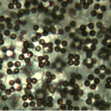
Phylum microspora (not to be confused with the green algae genus Microspora) contains some of the most unusual fungi: the microsporidia. There are over 1200 described species in this phylum (and that is only a fraction of their biodiversity), divided into about 150 genera (plural of genus). These organisms were originally thought to be protozoans, but recent DNA studies have demonstrated that they belong with the fungi. The microsporidia are all obligate parasites of animals and have an extremely reduced cell structure. They do not have mitochondria, so they can only grow and reproduce within the cells of their host. Their very resistant spores persist in the environment for a long time and allow them to spread from one animal to another. The spores are 1 to 40 micrometers long, making them the smallest eukaryotes. The spores are rougly oval and have a cell wall made of chitin that is thinnest at one end of the oval. Inside the cell membrane at this thinner end is an anchoring disc, which is connected to the most striking feature of microsporidian spores: the polar filament. The polar filament is a long, thin structure that is coiled around the inside of the spore from 5 to 15 times (depending on the species). Spores also have a vacuole, polaroplast, ribosomes, and one nucleus or two closely associated nuclei. Microspridia parasitize pretty much every animal group, from vertebrates to bryozoa (look that one up, they’re another weird form of life), but most are parasites of insects or other arthropods.
Microsporidia can have anywhere from a simple life cycle to a complex life cycle. The simplest life cycle follows the pattern: infect, produce spores. Complex life cycles may involve multiple hosts and multiple spore types. To infect a cell, osmotic pressure within the spore increases sharply and the polar filament is rapidly extruded from the spore through the process of eversion. In this process the filament is turned inside-out, similar to how you would turn a sleeve on a shirt inside-out. If the rapidly elongating filament contacts a cell, it will penetrate the membrane. Then the rest of the spore’s contents are injected into the host cell through the polar filament. The spore contents replicate more nuclei and begin to produce more spores. This continues until the host cell is completely filled with spores. The host cell then ruptures and releases the spores. Some species use an alternative method to enter the cell: they let the host cell eat them (phagocytosis). The spore contents are still extruded and they still produce spores, but they remain within a vacuole. Sometimes the parasites induce rapid cell division in the host cell, resulting in a xenoma. This is basically a tumor that allows the parasites to reproduce extremely quickly.
These tiny organisms do have a few impacts on humans. They have been investigated as a possible biological control agent for insects, and there is one species (Nosema locustae) that is commercially marketed to control grasshopper populations. Other species are economically significant pathogens of bees or silkworms. There are also at least 14 species that are known to infect humans and cause the disease microsporidiosis. Most of these are accidental or opportunistic pathogens that infect immuno-compromised individuals, however there are four species known to infect immuno-competent individuals. There has been a rise in microsporidiosis in recent years linked to the rise of HIV. Microsporidia may infect the gut, eye, muscles, or other parts of the human body.
See Further:
http://palaeos.com/eukarya/stem_metazoa/microsporidia.html
http://www.stanford.edu/group/parasites/ParaSites2006/Microsporidiosis/microsporidia1.html
http://wwx.inhs.illinois.edu/research/biocontrol/pathogens/typesofpathogens/microsporidia/

![#013: Characteristics of Phylum Basidiomycota [Archived]](https://www.fungusfactfriday.com/wp-content/themes/hueman/assets/front/img/thumb-medium-empty.png)


![#011: Characteristics of Kingdom Fungi [Archived]](https://www.fungusfactfriday.com/wp-content/themes/hueman/assets/front/img/thumb-small-empty.png)


1 Response
[…] disease caused by microsporidia, very simple pathogenic fungi that belong to the phylum Microspora (FFF#017), discovered that the tiny fungi can spread from one cell to the next by fusing together adjacent […]